Unlocking Biological Insight with the Human Anatomy 3D Model Pack 01
The visualization of complex biological systems has long been a cornerstone of medical education and scientific communication. Traditional methods, relying on two-dimensional textbooks or static cadaveric specimens, often struggle to convey the spatial relationships and dynamic nature of the human body. In an era where digital literacy is paramount, the shift toward immersive, interactive visual aids has become essential. The Human Anatomy 3D Model Pack 01 represents a significant step forward in this evolution, offering a comprehensive suite of high-fidelity assets designed to bridge the gap between abstract anatomical theory and tangible understanding. This collection is not merely a set of graphics; it is a toolkit for clarity, precision, and creative expression across diverse fields ranging from clinical training to digital entertainment.
Bridging the Gap Between Theory and Visualization
Understanding human anatomy requires more than memorizing names and functions; it demands a grasp of three-dimensional structure. How does the pancreas sit relative to the stomach? What is the precise curvature of the spine when viewed from different angles? These questions are difficult to answer with flat images. The Human Anatomy 3D Model Pack 01 addresses these challenges by providing models crafted with smooth shapes and vibrant colors that highlight distinct tissues and organ systems without overwhelming the viewer.
For educators and students alike, the ability to rotate, zoom, and isolate specific components is transformative. When a learner can mentally manipulate an object in space, retention rates improve significantly. The models in this bundle are designed with this cognitive process in mind. They strip away unnecessary clutter while maintaining anatomical accuracy, ensuring that the focus remains on the structural integrity and functional relationships of the organs. Whether examining the intricate folds of the brain or the branching pathways of the lungs, the visual fidelity allows for a deeper level of engagement that static media cannot provide.
Core Components of the Collection
The versatility of this resource stems from its breadth. The pack includes a wide array of essential human organs and body parts, meticulously rendered to serve as reliable references. Key elements include:
- Vital Organs: Detailed representations of the heart, liver, kidneys, stomach, intestines, and pancreas. These models capture the texture and form necessary for understanding physiological processes.
- Sensory Systems: High-quality illustrations of the eyes, ears, nose, and skin, allowing for close-up study of sensory reception and external barriers.
- Skeletal Framework: Critical structural elements such as the ribs, spine, pelvis, and teeth are included, providing a foundation for understanding movement and support.
- Reproductive and Urinary Systems: Comprehensive models that facilitate the study of these complex, often sensitive, biological systems with professional clarity.
Each element within the Human Anatomy 3D Model Pack 01 is carefully designed to look clean and modern. This aesthetic choice is deliberate; it ensures that the assets remain applicable in contemporary design contexts, from sleek medical presentations to engaging educational infographics. The vibrant color coding helps distinguish between muscle tissue, bone, and connective tissue, making complex anatomy easier to understand at a glance.
Workflow Integration: From Static Images to Dynamic Editing
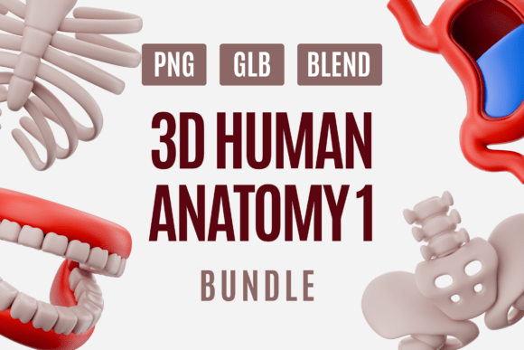
One of the most compelling features of this bundle is its multi-format availability. Understanding the needs of different users, the pack provides assets in PNG, GLB, and BLEND formats. This flexibility allows professionals to choose the level of interaction required for their specific projects.
For content creators who need quick turnaround times, the high-resolution PNG files offer immediate utility. These images are ready to be dropped into slide decks, blog posts, or marketing materials. They retain the crisp edges and vibrant coloring of the original 3D renders, ensuring that even in a flat format, the anatomical details remain sharp and legible. This is particularly useful for teachers preparing lesson plans or healthcare designers creating patient handouts where interactivity is not feasible but visual appeal is crucial.
However, for those requiring full control over the asset, the inclusion of GLB and BLEND files opens up a world of possibilities. The GLB format is optimized for web and mobile applications, allowing developers to embed interactive 3D models directly into websites or apps. Imagine a student using a tablet to peel back layers of the digestive system or a doctor showing a patient a rotating view of their own knee joint during a consultation. The lightweight nature of GLB ensures smooth performance without sacrificing quality.
For advanced users, the BLEND files invite deep customization. Using Blender or compatible 3D software, animators and designers can modify lighting, textures, and even the geometry of the models. Perhaps a project requires a specific cross-section of the heart or a stylized version of the brain for a sci-fi concept art piece. With access to the source files, the limitations are removed. This capability transforms the Human Anatomy 3D Model Pack 01 from a static library into a dynamic foundation for creative exploration.
Applications Across Diverse Industries
The utility of high-quality anatomical models extends far beyond the classroom. In the realm of medical education, these tools are indispensable. Medical students can use them to supplement their studies, visualizing structures that are difficult to observe in real-time dissections. Teachers can integrate these models into interactive whiteboards, turning passive lectures into active learning experiences. The ability to present the skeletal elements like the spine and pelvis in isolation helps clarify biomechanics and posture analysis.
In healthcare and clinical settings, clear communication is vital. Doctors and surgeons often rely on diagrams to explain procedures to patients. A well-rendered 3D model of the urinary organs or reproductive system can demystify complex conditions, reducing patient anxiety and improving informed consent. The clean, modern look of these assets ensures they fit seamlessly into professional presentations, enhancing the credibility of the information being conveyed.
Furthermore, the creative design and animation industries benefit immensely from accurate anatomical references. Character designers need to understand underlying musculature and bone structure to create believable movements in animated films or video games. Even if the final output is stylized, the foundational knowledge provided by these realistic models ensures that the resulting characters move and react in ways that feel organic and true to life. Animators can import the GLB files into their pipelines to rig characters or use the BLEND files as base meshes for further sculpting.
Enhancing Educational Outcomes Through Visual Clarity
The pedagogical value of the Human Anatomy 3D Model Pack 01 lies in its ability to simplify complexity. Cognitive load theory suggests that learners have limited capacity for processing new information. By presenting anatomical structures in a visually appealing and organized manner, these models reduce extraneous cognitive load, allowing students to focus on the core concepts.
Consider the challenge of teaching the relationship between the liver, gallbladder, and pancreas. Text descriptions can be dense and confusing. A 2D diagram might obscure depth. However, a 3D model allows the educator to show how these organs nestle together, how ducts connect, and how blood flow traverses the area. This spatial understanding is critical for future surgeons and biologists. The vibrant colors used in the pack help differentiate these structures instantly, reinforcing memory through visual association.
Moreover, the accessibility of these resources democratizes high-quality education. Not every institution has the budget for expensive physical models or cadaver labs. Digital assets like those found in this bundle provide a cost-effective alternative that scales easily. A single license can equip an entire department with the tools needed to visualize the wonders of the human body, from the smallest sensory receptors in the ear to the massive structural support of the pelvic girdle.
Future-Proofing Your Content Strategy
As technology advances, the demand for 3D content continues to grow. Virtual reality (VR) and augmented reality (AR) are becoming standard tools in medical training and public engagement. The Human Anatomy 3D Model Pack 01 is positioned perfectly to meet this demand. The GLB files are inherently compatible with many AR/VR platforms, meaning that today's assets can be tomorrow's immersive experiences with minimal conversion work.
For business owners and content strategists, investing in high-quality, versatile assets is a smart move. It ensures that your educational materials, marketing campaigns, or product demonstrations remain relevant as audiences shift toward interactive digital experiences. Whether you are creating an infographic about heart health or developing a training module for nursing staff, having access to editable, accurate 3D models gives you a competitive edge. It signals professionalism and a commitment to clarity.
Ultimately, the goal of any anatomical representation is to illuminate the intricate machinery of life. The Human Anatomy 3D Model Pack 01 achieves this by combining scientific rigor with artistic excellence. It empowers teachers to inspire, students to learn, designers to create, and professionals to communicate with unprecedented precision. By exploring the wonders of the human body through these detailed collections, we gain not just knowledge, but a deeper appreciation for the complexity and beauty of our own biology.